Ultrasound Physics & Machine Setting

About Course
What You Need to Know About Ultrasound Physics and Machine Settings
Ultrasound Physics and Machine Settings Before learning how to use an ultrasound machine, you should learn about the physics and general settings used in ultrasound machines. This will help you better understand how your machine works, leading to more accurate readings and more effective treatments. Here’s what you need to know about ultrasound physics and machine settings for the proper operation of your ultrasound machine.
Context
In the hands of an expert, the production of an ultrasound image looks easy. However, both image acquisition and interpretation are hugely reliant on a reasonable understanding of how ultrasound works. Where this is lacking, there is considerable scope for confusion and mis-di
Image Source: Pixabay
agnosis.
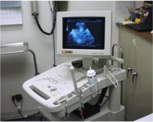
The basic components of an ultrasound system ultrasound machine are composed of a transducer to transmit and receive an ultrasound, a computer for storage and manipulation of the acquired data, and a monitor to display the image. Some form of image storage or hard copy device is needed to provide a permanent record of each examination.
The quality of the displayed data is determined by the operator’s skill and the inherent quality of each of the above components.
Generation and Detection of Ultrasound
Sound is simply the transfer of mechanical energy from a vibrating source through a medium, for example, the plucking of a guitar string makes the string vibrate and produces a sound.
Ultrasound is defined as the sound of a frequency above the human audible range, i.e. above 20 kHz. For most emergency, medicine applications frequencies in the region of 1 to 10 MHz are used.
An ultrasound transducer (image, right) converts electrical energy into a mechanical pulse of sound that is transmitted to the patient. Returning echoes are converted into an electrical signal from which the image is formed.
The Pulse Echo Principle
Echo-location
Medical ultrasound utilizes the pulse-echo principle to construct a two-dimensional image of anatomical structures within a patient.
This is essentially the same principle used by bats to catch insects through echolocation.
Ultrasound is not emitted continuously from the transducer, as there would be no way of knowing how long a wave had been traveling once it returned to the transducer. Instead, transducers oscillate between emitting a pulse of ultrasound, and then in the pause that follows, the time that elapses for the echoes to be detected is measured.
Acoustic Impedance
A pulse of sound leaving the transducer will travel into the patient until it encounters a change in acoustic impedance (Z) (Fig 1). Acoustic impedance refers to the resistance of the tissue due to molecular movement. It is directly related to tissue density
At such an interface, a proportion of the sound energy is reflected back to the transducer and this returning echo is detected. If the speed of sound is known and the time taken for the echo to return is measured, the depth of the reflecting interface can be calculated:
Types of Ultrasound
Each pulse of sound transmitted to the patient will generate a stream of returning echoes from multiple reflecting interfaces at various depths within the tissue. Although all ultrasound is based on reflected sound, the means by which the echoes are analyzed and displayed varies. This gives rise to several types, or modes, of ultrasound:
- A-Mode
- B-Mode
- M-Mode
A-mode
A mode is short for amplitude modulation.
A-mode displays as a graph on the screen with the x-axis as time, and the y-axis as amplitude. It is used in scanning the eye. The diagram shows an ultrasound of the abdomen with the A-mode display corresponding to each layer.
Course Content
1. Ultrasound Class of Physics & Machine Settings
-
Lecture 1 | Ultrasound Class of Physics & Machine Settings
31:10